← Back to home

Minimally invasive ankle surgery

Ankle arthroscopy

Visualising and treating the inside of the ankle joint through small incisions using a high-definition camera and miniature instruments.

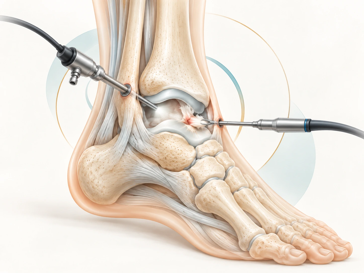
Medical illustration of ankle arthroscopy

What is ankle arthroscopy?

Arthroscopy involves introducing a small camera — the arthroscope — into the ankle. It provides direct visualisation of cartilage, synovium, intra-articular ligaments and mechanical lesions.

Through a second small portal, instruments can remove inflamed tissue or loose bodies, resect bony impingement, or treat selected cartilage lesions. Arthroscopy can also accompany other stabilisation or reconstructive procedures.

What it is notArthroscopy is not simply a 'camera to have a look'. It should answer a specific clinical indication and enable a therapeutic procedure likely to improve symptoms.
Indications

Which conditions can be treated?

Anterior impingement

Pain at the front of the ankle, often worse in dorsiflexion, related to osteophytes and/or abnormal soft tissue.

Osteochondral lesion

Localised injury to the cartilage and subchondral bone of the talus. Treatment depends on lesion size, depth and stability.

Loose bodies & synovitis

Mobile fragments or abnormal synovial tissue that may cause pain, swelling, catching or locking.

Posterior impingement / os trigonum

Posterior pain, particularly in forced plantar flexion. Posterior arthroscopy provides access to this region.

Instability & ligament injuries

Chronic ligament injuries may cause pain, giving way and instability. Depending on the lesion, treatment may combine arthroscopy with ligament repair or reconstruction.

Achilles tendon

Insertional or mid-portion tendinopathy and selected ruptures may require tailored surgery, sometimes minimally invasive, but not isolated ankle arthroscopy.

Peroneal tendons

Peroneal tendinopathy, tears, ruptures or instability may cause lateral ankle pain. Treatment is tailored to the lesion and may be combined with treatment of ligament instability.

Arthritis: an important distinction

Arthroscopy may be useful when focal impingement or a loose body explains part of the symptoms. However, simple arthroscopic 'clean-up' is not a reliable treatment for diffuse advanced arthritis; other strategies should then be considered.

Technique

What happens during surgery?

Plan

Clinical examination is correlated with radiographs and, depending on the condition, MRI or CT. The aim is to define the cause of symptoms before surgery.

Access the joint

Small incisions — called portals — are made while protecting nearby nerves, vessels and tendons.

See & treat

The camera explores the joint. Miniature instruments then perform the planned procedure: targeted debridement, osteophyte resection, loose-body removal or cartilage treatment.

Tailor recovery

Weight-bearing, immobilisation and rehabilitation depend heavily on the procedure performed. Simple impingement resection and cartilage repair do not have the same protocol.

Goals

Why choose an arthroscopic approach?

Direct vision

Precise inspection of intra-articular structures and targeted treatment of the lesion.

Small incisions

Less soft-tissue exposure than with some open approaches.

Targeted recovery

For simple procedures, recovery can be relatively quick; cartilage repair or associated procedures require greater protection.

Recovery

Recovery depends mainly on what was treated

There is no single ankle arthroscopy protocol. After debridement or impingement resection, weight-bearing can sometimes resume quickly. After cartilage repair or an associated procedure, a period of protected or non-weight-bearing may be required.

First daysControl pain & swelling

Elevation, ice as instructed, pain medication and ankle protection.

1–2 wksWound healing

Review of the small incisions and adjustment of weight-bearing and motion.

Following weeksMotion & strength

Progressive rehabilitation according to the condition and procedure performed.

SportIndividualised return

Timing varies substantially and depends on the lesion, any cartilage procedure, stability and the sport involved.

Risks

What are the risks?

Arthroscopy remains a surgical procedure. Complications are uncommon but include infection, wound problems, thrombosis, persistent swelling or stiffness, cartilage injury, and irritation or injury to a cutaneous nerve near a portal. Exact risk also depends on associated procedures.

Key pointSuccess depends less on incision size than on diagnostic accuracy and appropriate indication. Ankle pain without a clearly identified cause is not, by itself, an indication for arthroscopy.
FAQ

Frequently asked questions

Is it day surgery?

Many isolated arthroscopies can be performed as day surgery, but this depends on the procedure, anaesthesia, associated surgery and the patient's medical situation.

Is MRI always required?

No. Imaging depends on the suspected problem. Weight-bearing radiographs remain essential in many situations; MRI or CT is added when it provides useful information for decision-making.

Can arthritis be treated with arthroscopy?

A focal impingement associated with arthritis can sometimes be treated. In diffuse advanced arthritis, however, arthroscopic debridement does not restore cartilage and its benefit is limited.

Selected medical sources

General patient information; it does not replace individual consultation and diagnosis.

  1. Glazebrook MA et al. Evidence-based indications for ankle arthroscopy. Arthroscopy. 2009.
  2. Epstein DM et al. Anterior ankle arthroscopy: indications, pitfalls, and complications. Foot Ankle Clin. 2015.
  3. Leiber-Wackenheim F. Anterior ankle impingement. Orthop Traumatol Surg Res. 2025.
  4. Vega J et al. Posterior ankle and subtalar arthroscopy: indications, technique, and results. 2012.
  5. Ankle Arthroscopy. StatPearls. Updated 2026.

Helping you move forward, step by step.

A consultation is an opportunity to discuss your situation, goals and the different treatment options.

Specialist consultation

Persistent ankle pain?

The aim of consultation is to identify the source of pain precisely and determine whether arthroscopy, non-operative treatment or another procedure is the most appropriate strategy.